The skull base is the floor of the skull, the bony shelf on which the brain rests. Important nerves for vision, smell, eye movement and facial feeling pass through it, along with major arteries. Tumours here used to mean large openings in the skull. For many of them, especially pituitary tumours, the route today is through the nose using an endoscope.
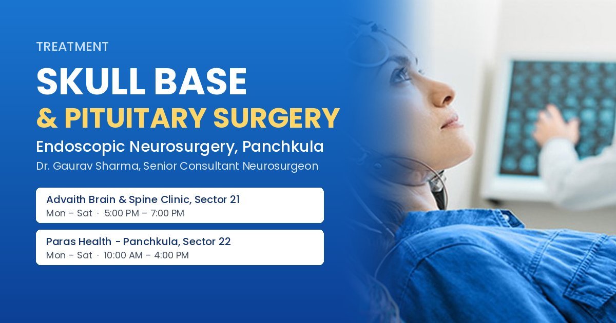
Dr. Gaurav Sharma, Senior Consultant Neurosurgeon in Panchkula, performs endoscopic skull base and pituitary surgery as part of his brain tumour work. Whether this approach suits a particular tumour depends on its size, its spread and what it is attached to.
An endoscope is a thin tube with a light and a high-definition camera. It is passed through the nostril to the sphenoid sinus at the back of the nose, which sits right below the pituitary gland. A small window is made in the bone, and the tumour is removed with fine instruments while the surgeon watches on a screen. There is no cut on the face or scalp.
Because the brain does not need to be lifted to reach the tumour, many patients recover faster than after open surgery. That said, every tumour is different, and some still need a craniotomy or a combination of approaches.
The pituitary is a pea-sized gland that controls many of the body's hormones. Most pituitary tumours (adenomas) are benign, but they can cause trouble in two ways.
Not all pituitary tumours need surgery. Prolactin-producing tumours, for example, often shrink with tablets alone. An endocrinologist is usually involved in the decision.

The endoscopic route may also be used for some meningiomas at the front of the skull base, craniopharyngiomas, Rathke's cleft cysts and leaks of brain fluid (CSF) through the nose. Larger or more spread-out tumours may still need open surgery, which is explained on the brain tumour surgery page.
Expect an MRI with contrast, blood tests for pituitary hormones, and an eye check including visual fields. A CT scan helps plan the route through the sinuses. Bring all old scans and reports, as comparing them shows whether the tumour has grown.
Most patients stay in hospital for a few days. A blocked nose and crusting are common for a few weeks; nose-blowing, straining and heavy lifting are avoided while the repair heals. Hormone levels and urine output are watched closely, since the pituitary may need time to settle, and some patients need hormone tablets for a while or longer. Vision often improves when pressure on the nerves is relieved, though this cannot be promised for everyone.
Follow-up MRI scans are done at intervals to check for any remaining or returning tumour.
Skull base tumours grow slowly and their early signs are easy to put down to other causes. Gradual loss of side vision, headaches behind the eyes, unexplained hormone problems such as missed periods or milk discharge, a constantly blocked nose on one side with reduced smell, or clear watery fluid dripping from the nose (especially on bending forward) are all reasons to see a doctor and consider an MRI. Many of these turn out to be something simple, but when a tumour is found early, the options are usually wider and the surgery more straightforward.
| Location | Days | Timings |
|---|---|---|
| Advaith Brain & Spine Clinic, 1842, Sector 21, Panchkula – 134112 | Monday – Saturday | 5:00 PM – 7:00 PM |
| Paras Health - Panchkula, near Nada Sahib Gurudwara, Sector 22, Panchkula – 134109 | Monday – Saturday | 10:00 AM – 4:00 PM |
| Panchkula OPDs | Sunday | By appointment |
Phone / WhatsApp: +91-9780703819
No visible scar. The surgery is done through the nostril. Occasionally a small piece of tissue is taken from the thigh or nose lining for the repair.
Some people notice a reduced sense of smell for a few weeks while the nose heals. Lasting change is uncommon.
Some can, which is why follow-up MRI scans and hormone tests continue for years after surgery.
No. Size, spread and the surrounding nerves and vessels decide the approach. Some tumours need open or combined surgery.